3 Minute Read
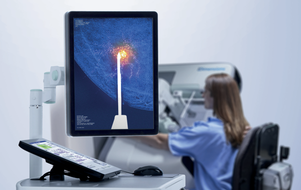

In breast imaging, catching subtle abnormalities early is everything. Contrast-Enhanced Mammography (CEM) has transformed how we spot tricky or non-calcified breast lesions, offering exceptional sensitivity. However, as the use of CEM grows, clinical teams face a familiar hurdle: how to quickly and accurately sample a lesion that is only visible under contrast.
Contrast-guided biopsies bridge this critical gap, allowing healthcare teams to target and sample contrast-enhancing lesions directly on the mammography system, without the delays, high costs, or scheduling bottlenecks of alternative imaging.
From Detection to Diagnosis: How It Works
Traditionally, if an abnormality was visible only via contrast enhancement (like on a breast MRI or CEM) but couldn’t be located on ultrasound or standard 2D mammography, biopsy options were limited. Patients often had to wait for expensive MRI-guided biopsy slots or undergo complex correlation attempts.
Contrast-guided biopsy simplifies this pathway into a smooth, step-by-step process:
The entire procedure can be completed in 15 to 20 minutes, compared to 45 minutes or longer for an MRI-guided alternative. For patients, this means less time in compression, a comfortable seated position, and significantly less anxiety.
The Diagnostic Foundation: I-View Contrast-Enhanced Imaging
Before a targeted contrast biopsy can take place, clinicians need a fast, reliable way to identify vascular abnormalities. This is where I-View Contrast-Enhanced Imaging from Hologic plays a vital role.
Working seamlessly on Hologic 3D Mammography™ systems, I-View™ software combines functional contrast imaging with traditional mammography in a single, short exam. By subtracting background dense tissue clutter, I-View™ allows clinical teams to spot subtle, hyper-vascularised lesions that might otherwise remain hidden on 2D mammograms or ultrasound, delivering diagnostic sensitivity comparable to breast MRI right within the mammography suite.
Unlocking a Seamless Workflow: Affirm Contrast Biopsy Software
When an abnormality is discovered using I-View CEM, having a direct pathway to sampling is essential. To help clinics and trusts deliver this complete continuum of care, Hospital Services Limited (HSL) is proud to supply the Affirm® Contrast Biopsy Software alongside I-View™ for Hologic systems.
By integrating directly with Hologic 3D Mammography gantries, the Affirm Contrast Biopsy Software transforms diagnostic units into fully equipped interventional suites.
Clinical teams choose Affirm Contrast because it allows them to confidently target subtle, vascular-only lesions that standard 2D mammography, tomosynthesis, or ultrasound frequently miss. By enabling a seamless transition from diagnostic imaging to targeted biopsy within the exact same clinical visit, it significantly shortens the patient journey from initial discovery to a clear treatment plan. Additionally, bringing contrast biopsy directly into the mammography department alleviates the heavy strain on overbooked MRI suites, while fast image acquisition keeps compression times to a minimum and maintains exceptionally stable target positioning for maximum patient comfort.
Elevating Patient Outcomes with HSL
As Contrast-Enhanced Mammography rapidly expands across healthcare institutions, having an integrated route from initial screening (I-View) to histological diagnosis (Affirm Contrast) is essential. Together, these technologies ensure that every enhancement identified can be evaluated and sampled promptly, giving clinicians clarity and patients fast, reliable answers.
To learn more about incorporating I-View Contrast-Enhanced Imaging and the Affirm Contrast Biopsy Software into your facility’s imaging suite, get in touch with the HSL team today.







 Back to All News